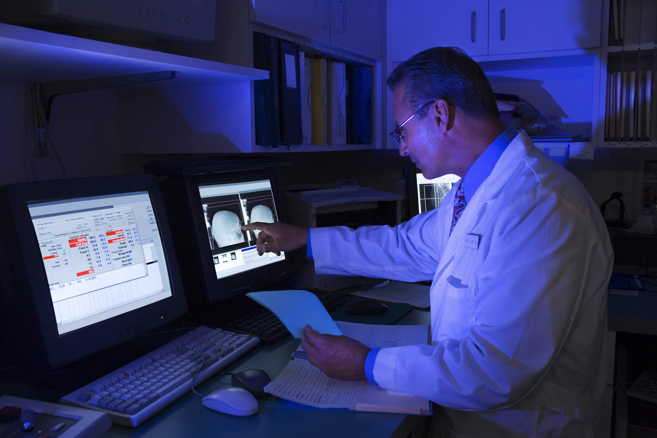
A machine-learning approach to analysing lung cancer tissue is more reliable than the opinion of even a highly experienced pathologist, suggests a new study by the Stanford University School of Medicine. Its authors believe that such an approach would be effective in treating other forms of cancer, too.
Krista Conger of the medical school’s Office of Communication & Public Affairs described the four key actions of the study’s researchers:
- The analysis of 2,186 adenocarcinoma and squamous-cell-carcinoma images provided by The Cancer Genome Atlas, a national database that features “information about the grade and stage assigned to each cancer and how long each patient lived after diagnosis.”
- The training of software “to identify many more cancer-specific characteristics than can be detected by the human eye — nearly 10,000 individual traits, versus the several hundred usually assessed by pathologists. These characteristics included not just cell size and shape, but also the shape and texture of the cells’ nuclei and the spatial relations among neighbouring tumour cells.”
- The homing in on “a subset of cellular characteristics identified by the software that could best be used to differentiate tumour cells from the surrounding noncancerous tissue, identify the cancer subtype, and predict how long each patient would survive after diagnosis.”
- The validating of the software’s ability to “accurately distinguish short-term survivors” from nearly 300 survivors tracked in a separate dataset, all of whom lived notably longer.
“The work,” writes Conger, “is an example of Stanford Medicine’s focus on precision health, the goal of which is to anticipate and prevent disease in the healthy and precisely diagnose and treat disease in the ill.”
Read the full article here.
Orion Health built a platform that plugs the gap between HIEs and analytics systems. Learn more: read the Chilmark Research report now!


